Home
Uncategories
Left Hip Muscles Anatomy - Lower Back Pain | The Hip-Flexor Fix - Therapeutic Personal Trainers North Vancouver : Your email address will not be published.
Left Hip Muscles Anatomy - Lower Back Pain | The Hip-Flexor Fix - Therapeutic Personal Trainers North Vancouver : Your email address will not be published.
Left Hip Muscles Anatomy - Lower Back Pain | The Hip-Flexor Fix - Therapeutic Personal Trainers North Vancouver : Your email address will not be published.. These muscles constitute the anatomical classification known as the medial compartment of the thigh. Through a simple and intuitive interface it is possible to observe every anatomical structure from any angle. Each muscle below has the bones in bold for intermediate learners and the specific bony landmarks for advanced learners. Knee assessment and hip mechanics learn how hip and pelvis mechanics can influence the knee powered by physiopedia start course. Now that you watched the video, you.
Most modern anatomists define 17 of these muscles, although some additional muscles may sometimes be considered. Knee assessment and hip mechanics learn how hip and pelvis mechanics can influence the knee powered by physiopedia start course. The cavity of the acetabulum the external obturator muscle is short external rotator muscle of hip joint. The muscular system is responsible for the movement of the human body. One example of an ab exercise that actually focuses.
File:Anterior Hip Muscles 2.PNG - Wikimedia Commons from upload.wikimedia.org The hip's unique anatomy enables it to be both extremely strong and amazingly flexible, so it can bear weight and allow for a wide range of movement. In order to isolate the abdominals, you need to minimize the involvement of the hip flexors and maximize the contraction of the abdominals. Pelvis and acetabulum, with muscle attachment sites. These muscles are responsible for hip joint extension (backward movement). The muscles of the neck can be divided into groups according to their location. In conclusion, a thorough understanding of pelvic and hip anatomy is important for. This anatomical atlas was especially designed for a specific public (radiologists, surgeons, rheumatologists and physicians specializing in musculoskeletal imaging). Highly detailed 3d models, with textures up to 4k resolution, enable to examine the shape of each.
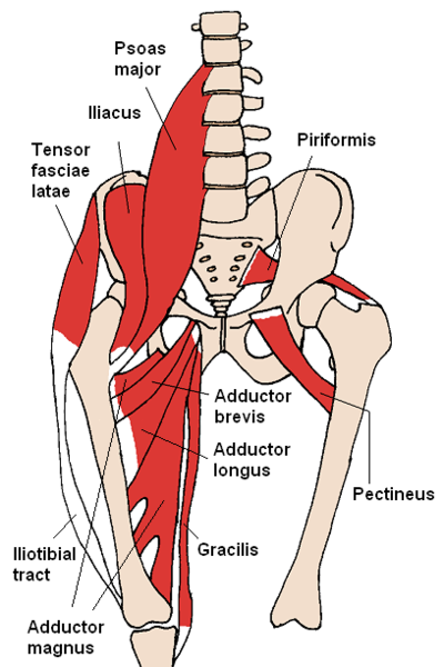
936 x 504 png 317 кб.
It originates at the anterior inferior iliac spine and just above the acetabulum of the hip bone. Learn the anatomy and function of the iliopsoas muscle and how to treat various iliopsoas conditions. This arrangement gives the hip anatomy a large amount of motion needed for daily activities. The psoas major muscle (usually shortened to just the psoas muscle) is one of the muscles of the posterior abdominal wall and lies not in the retroperitoneum but posterior to it, in the iliopsoas compartment. The cavity of the acetabulum the external obturator muscle is short external rotator muscle of hip joint. Anatomy of a human body we study anatomy. There are a lot of muscles of the hip and thigh. Each muscle below has the bones in bold for intermediate learners and the specific bony landmarks for advanced learners. Back muscles of the hip. It is a flat, triangular muscle on the anterior wall of the pelvis. The iliopsoas muscle is a major hip flexor. The hip's unique anatomy enables it to be both extremely strong and amazingly flexible, so it can bear weight and allow for a wide range of movement. In clinical anatomy the thigh muscles are divided into three groups:
Muscle movements, types, and names. If you know all the hip flexor names and bones they attach to, that's an awesome accomplishment! Knee assessment and hip mechanics online course: A bursa that sometimes causes problems in the hip is sandwiched between the bump on the outer hip (the greater trochanter) and the muscles and tendons that cross over the bump. 3 months later i got acute excrutiating pain in inguinal area.
MRI anatomy of hip joint | free MRI axial hip anatomy from mrimaster.com Learn about hip muscles human anatomy with free interactive flashcards. The cavity of the acetabulum the external obturator muscle is short external rotator muscle of hip joint. The hip's unique anatomy enables it to be both extremely strong and amazingly flexible, so it can bear weight and allow for a wide range of movement. Anatomy of a human body we study anatomy. These muscles are responsible for hip joint extension (backward movement). Back muscles of the hip. The iliopsoas muscle is a major hip flexor. for detailed anatomy of pelvic bones, read anatomy of hip bone.
Anterior muscles extend your legs and flex your thighs.
Knee assessment and hip mechanics learn how hip and pelvis mechanics can influence the knee powered by physiopedia start course. The muscles of the neck can be divided into groups according to their location. One example of an ab exercise that actually focuses. Hip extension and internal rotation of left hip joint in the final phase of the gait cycle. Now that you watched the video, you. These muscles work together to flex your hip and to stabilize your hip and lower back during activities such as walking, running, and rising from a chair. Leave a reply cancel reply. A bursa that sometimes causes problems in the hip is sandwiched between the bump on the outer hip (the greater trochanter) and the muscles and tendons that cross over the bump. I pulled some muscles on left hip hiking. Learn their anatomy efficiently and easily using kenhub's muscle anatomy and reference charts! Through a simple and intuitive interface it is possible to observe every anatomical structure from any angle. Rectus femoris forms the middle portion of the quadriceps. Muscles that act on the lower limb cause movement at the hip, knee and foot joints.
The muscles of the neck can be divided into groups according to their location. The hip's unique anatomy enables it to be both extremely strong and amazingly flexible, so it can bear weight and allow for a wide range of movement. Anatomy 3d atlas allows you to study human anatomy in an easy and interactive way. The iliopsoas muscle is a major hip flexor. Learn about hip muscles human anatomy with free interactive flashcards.
Hip Muscles - The lateral rotators are muscles whose main job is basically what it sounds like ... from i.pinimg.com The iliopsoas muscle is a major hip flexor. The hip muscles encompass many muscles of the hip and thigh whose main function is to act on the thigh at the hip joint and stabilize the pelvis. Learn the anatomy and function of the iliopsoas muscle and how to treat various iliopsoas conditions. Rectus femoris muscle, one of the quadriceps muscles on the front of your thigh. The psoas major muscle (usually shortened to just the psoas muscle) is one of the muscles of the posterior abdominal wall and lies not in the retroperitoneum but posterior to it, in the iliopsoas compartment. Most modern anatomists define 17 of these muscles, although some additional muscles may sometimes be considered. Hip muscle strains can improve with home treatment, but severe strains may need physical therapy and possibly even surgery. The hip joint is the articulation of the pelvis with the femur, which connects the axial skeleton with the lower extremity.
for detailed anatomy of pelvic bones, read anatomy of hip bone.
Each muscle below has the bones in bold for intermediate learners and the specific bony landmarks for advanced learners. It is ideal for classrooms or doctor's offices, and. Rectus femoris forms the middle portion of the quadriceps. The psoas major muscle (usually shortened to just the psoas muscle) is one of the muscles of the posterior abdominal wall and lies not in the retroperitoneum but posterior to it, in the iliopsoas compartment. Microscopic anatomy of skeletal muscle. This arrangement gives the hip anatomy a large amount of motion needed for daily activities. The muscles of the neck can be divided into groups according to their location. One example of an ab exercise that actually focuses. These muscles are responsible for hip joint extension (backward movement). Learn their anatomy efficiently and easily using kenhub's muscle anatomy and reference charts! The muscles of the pelvis, hip and buttock anatomical chart shows how each muscle in this area of the body works with the others, and the you will not find a more comprehensive or more detailed examination of these muscles in an anatomy chart. Anterior muscles extend your legs and flex your thighs. Through a simple and intuitive interface it is possible to observe every anatomical structure from any angle.


0 Comments:
Posting Komentar